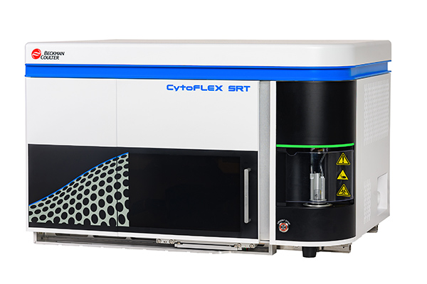
Fluorescence activated cell sorting
Technology / Methodology: 
Cell characterization
Location:
CEITEC MU, building C04, room 2.19
Research group:
CF: Core Facility Biomolecular Interaction and Crystallography
Fluorescence-activated cell sorting (FACS) is an enhanced form of flow cytometry that utilizes fluorescent markers to sort and analyze cells, allowing researchers to accurately isolate specific cell populations.
The CytoFLEX SRT is a compact cell sorter designed to handle a wide range of samples commonly processed in a core facility. From bacteria to tumor cells and everything in between, the SRT provides the versatility needed to support the diverse research community.
Features:
2 lasers (488nm and 638nm) with 5 channels

The FACS Process:
FACS protocols generally consist of four key phases:
- Sample Preparation and Labeling: Researchers start by preparing the cell sample, ensuring the cells are viable and evenly suspended. Fluorescent labels, typically dyes attached to antibodies, are introduced to the sample, binding to specific surface markers unique to each cell type.
- Laser Excitation and Cell Interrogation: Once labeled, cells are passed through the flow cytometer individually. As they encounter a laser beam, the fluorescent tags are excited, causing each cell to emit light at different wavelengths based on the labels they carry.
- Signal Detection and System Analysis: Advanced detectors within the FACS system capture both the emitted fluorescence and scattered light from the cells. Forward scatter provides data on cell size, while side scatter gives insights into the cell’s granularity or internal complexity. The fluorescence reveals the presence and quantity of specific cell markers based on the fluorescent tags bound to the cells.
- Cell Sorting and Collection: After detection, cells are electrically charged and pass through an electromagnetic field within the sorter. This field deflects the cells into separate containers based on their charge, effectively sorting them according to their predetermined fluorescence profiles.
Measurement on CytoFlex is supposed to be performed manually by user itself after training.
For more details, please contact responsible person:
Eva Paulenová
e-mail: eva.paulenova@ceitec.muni.cz
phone: +420 549 49 78 22
office: 3.37/C04


